

CLINICAL SOLUTION / ENT
Ear & Nasal Examination
Start with
the anatomy.
The ear canal is curved and delicate, leading to the tympanic membrane and middle ear.
The nasal cavity is longer and more variable, with turbinates and meatuses that require flexible navigation.
Different anatomy. Different approach. Better outcomes.
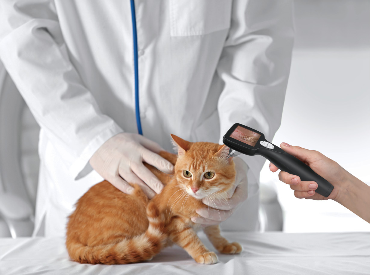
Two pathways.
One clear decision.
EAR EXAMINATION
01
Patient factors
Assess size, ear conformation and cooperation.
02
Target anatomy
Define outer ear, ear canal or middle ear.
03
Access and instrumentation
Select the scope and tools that fit the canal.
NASAL EXAMINATION
01
Patient factors
Assess size, respiratory status and tolerance.
02
Target anatomy
Identify nares, nasal cavity, meatuses or sinus.
03
Access and instrumentation
Choose scope diameter and working channel for the task.
What defines
the setup?
♧
Patient size
Match scope and access to patient anatomy.
⊙
Target anatomy
Clarify structures and clinical objectives.
⌀
Access diameter
Balance visibility with patient comfort.
⌁
Sampling or retrieval need
Confirm channel size and instrument compatibility.
Visibility is only
the beginning.
Stable handling. Clear documentation. Efficient workflow. The right pathway supports diagnosis, treatment and follow-up.
Relevant products.
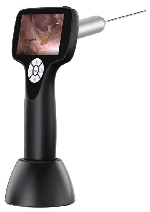
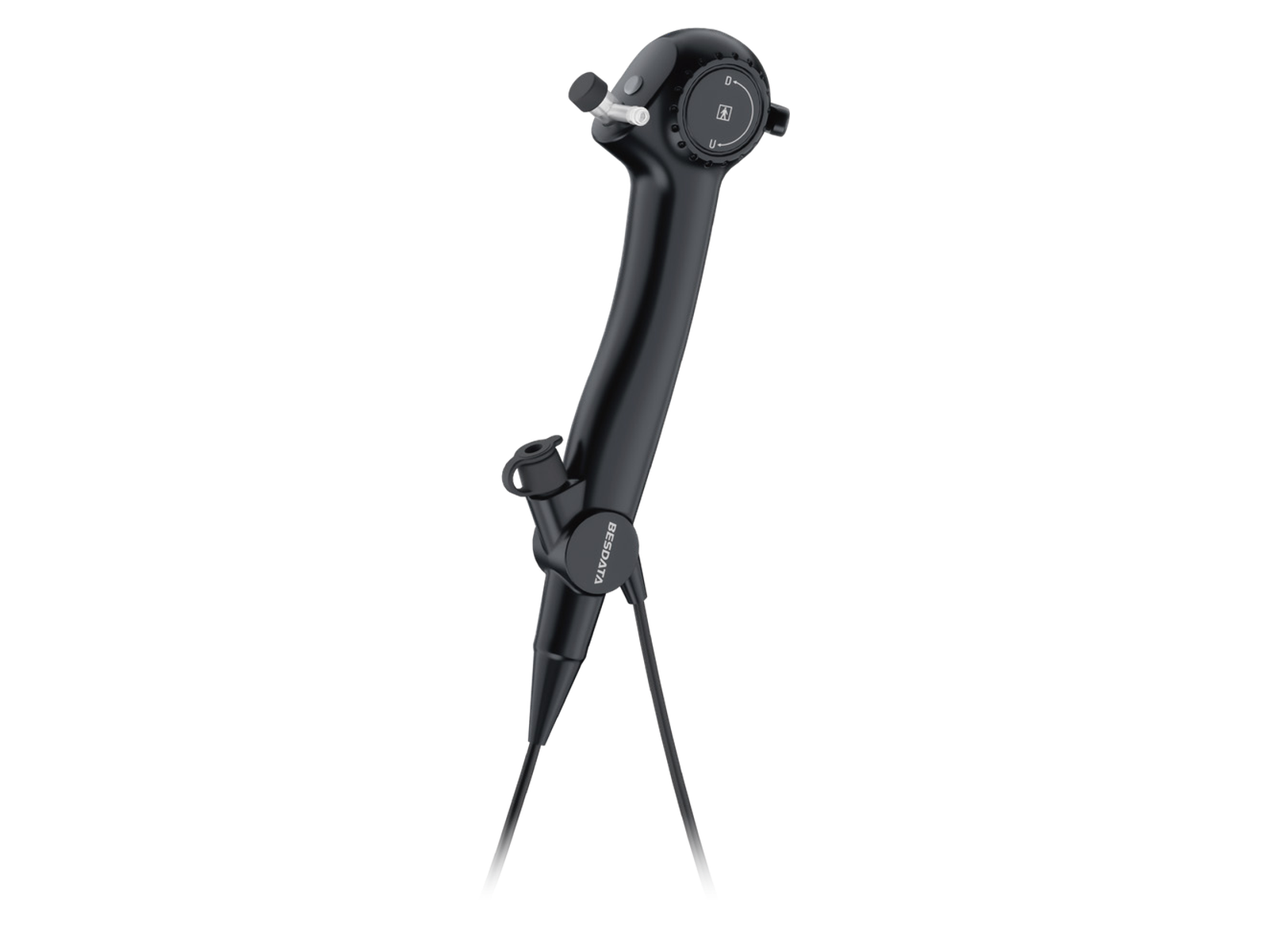
Veterinary Flexible Endoscope
Flexible access, clear imaging and versatile clinical use.