

CLINICAL SOLUTION / URINARY
Urinary Endoscopy
Small anatomy.
Precise access.
Urinary endoscopy requires careful alignment between patient anatomy, access route and scope diameter.
The intended examination may involve visualization alone or require irrigation, retrieval, sampling or compatible instruments.
Define the target. Protect access. Configure the system around the procedure.
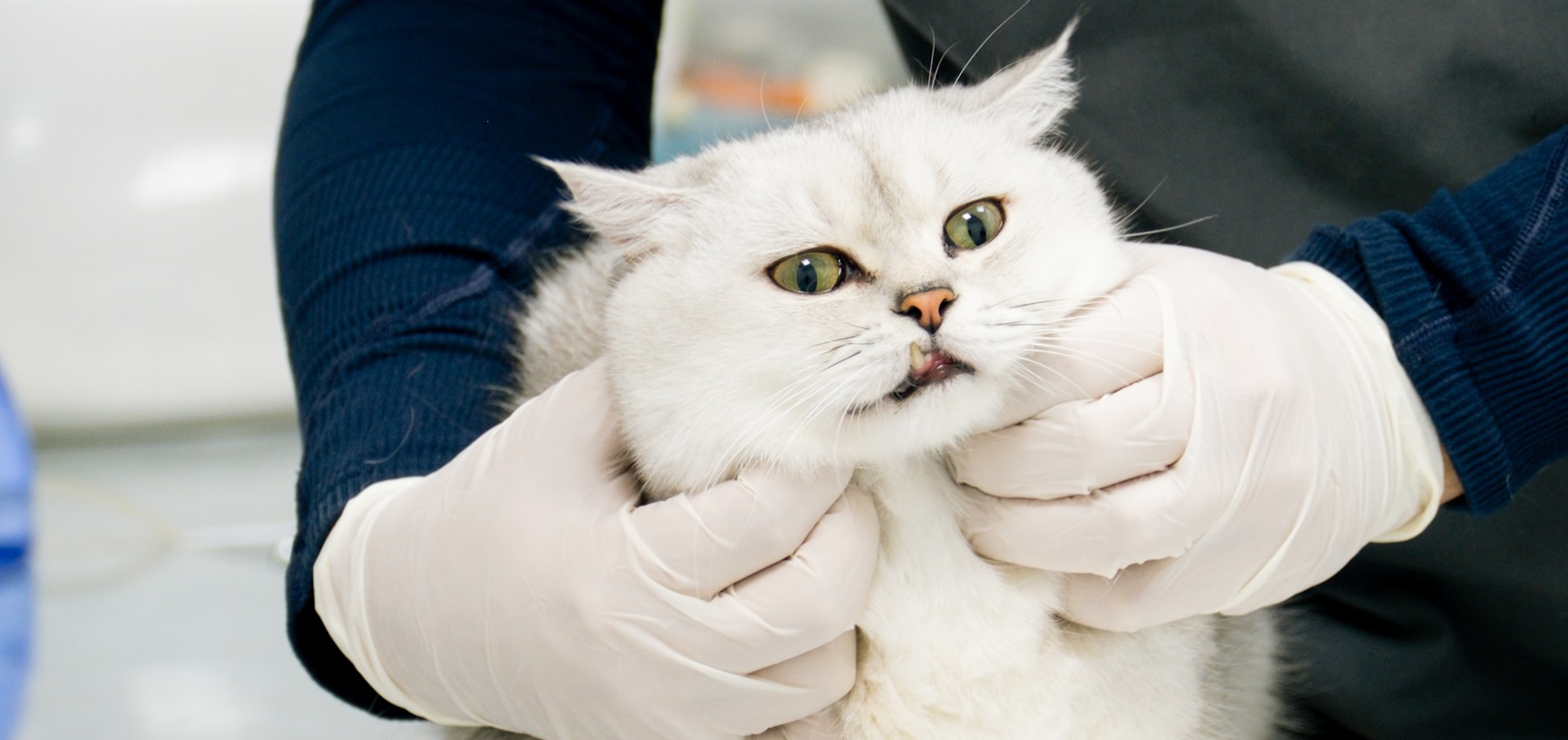
Inspection and intervention.
Plan both from the start.
LOWER URINARY ACCESS
01
Patient anatomy
Consider size, sex and the intended access route.
02
Target area
Define urethral, bladder or adjacent visualization.
03
Scope selection
Match diameter, flexibility and field of view.
TARGETED PROCEDURES
01
Working channel
Confirm instrument and irrigation compatibility.
02
Procedure objective
Plan sampling, retrieval or guided intervention.
03
System coordination
Align imaging, accessories and fluid management.
What shapes
urinary access?
♧
Patient anatomy
Select access around size and route.
⊙
Scope diameter
Balance access, visibility and handling.
⌀
Fluid workflow
Plan irrigation and image clarity.
⌁
Instrument need
Confirm channel and accessory compatibility.
Precision depends
on the complete setup.
The scope, imaging platform, fluid pathway and instruments should be planned as one urinary endoscopy workflow.
Relevant products.
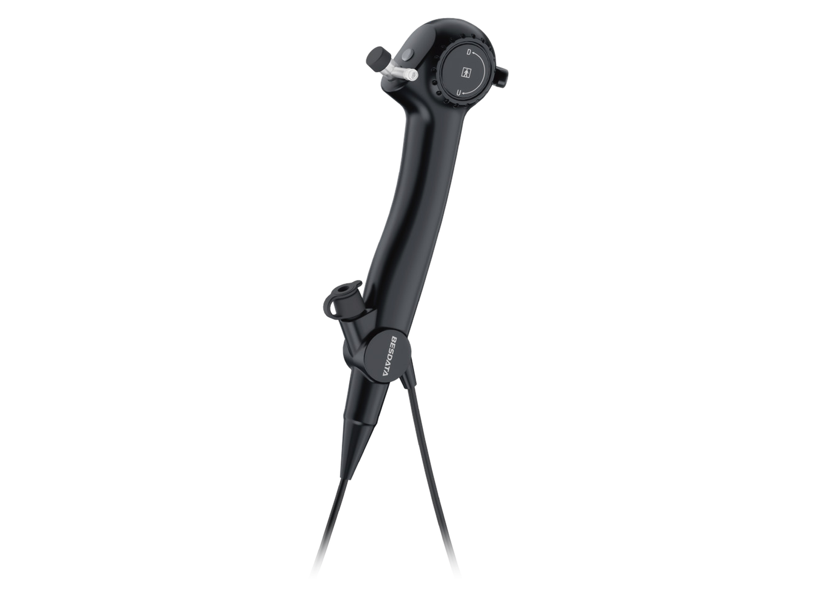
Veterinary Flexible Endoscope
Flexible visualization for urinary access and minimally invasive examination.
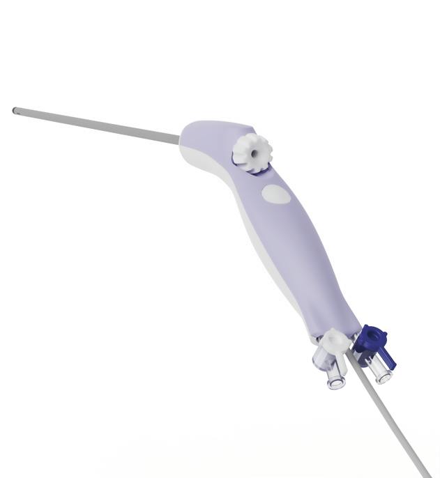
Veterinary Rigid Endoscope
Rigid visualization and accessory compatibility for selected urinary procedures.