

CLINICAL GUIDE
Upper GI endoscopy in dogs and cats.
A practical guide to clinical scope, system configuration, view management and a repeatable examination workflow.
8 MIN READ / CLINICAL GUIDE / AUGUST 2026
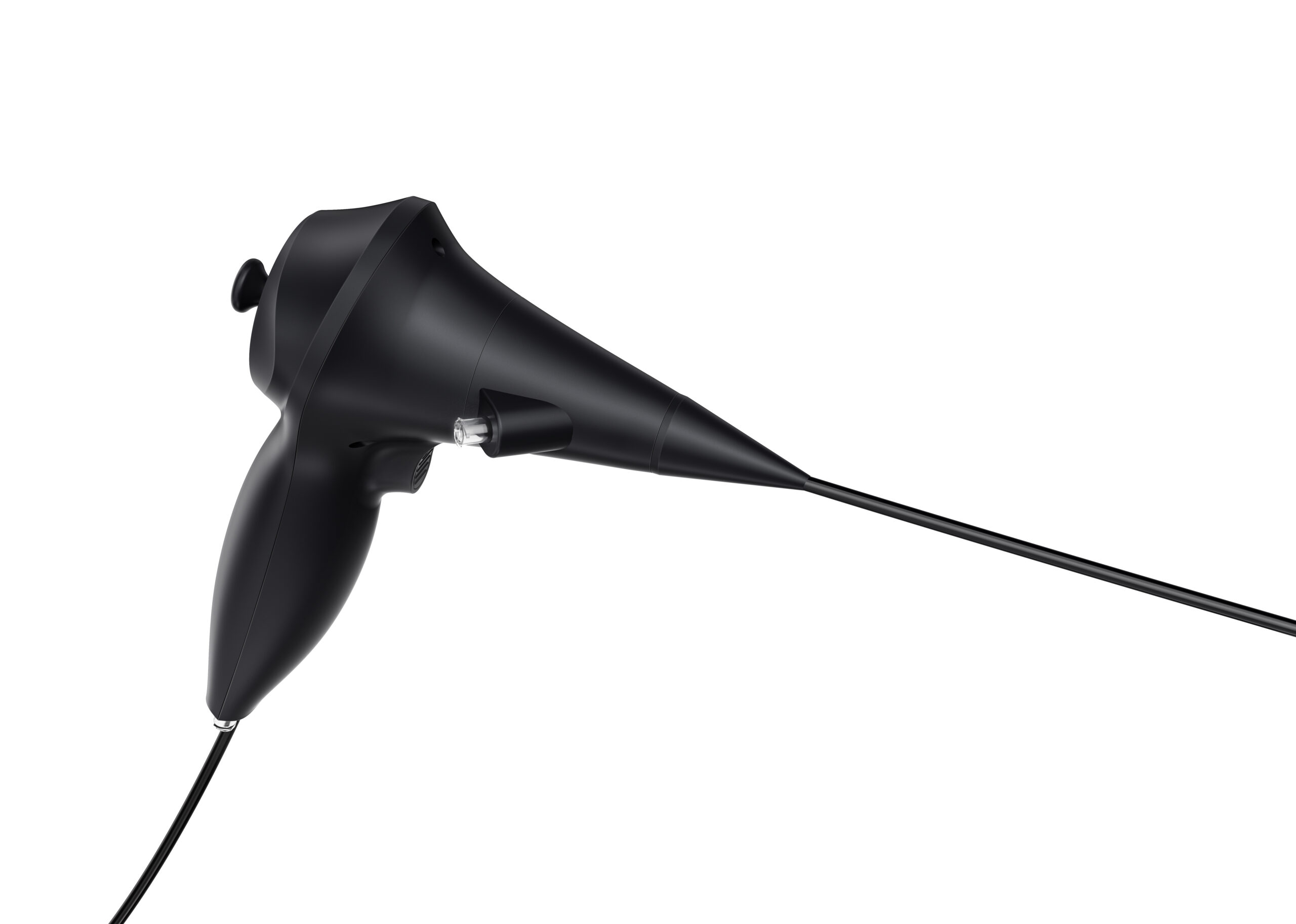
Upper gastrointestinal endoscopy gives veterinary teams a direct view of the mucosal surface of the esophagus, stomach and accessible proximal duodenum. It can support lesion documentation, targeted sampling and selected interventional procedures when clinically appropriate.
The examination is one part of a wider diagnostic pathway. Patient history, physical examination, laboratory findings and diagnostic imaging still guide whether endoscopy is appropriate and what the team should be prepared to do during the procedure.
KEY POINT
Start with the clinical question and patient anatomy. Scope diameter, working length, angulation, channel size and view-management functions should follow the case—not the other way around.
What upper GI endoscopy can examine.
A flexible veterinary gastroscope can provide visual access to several upper gastrointestinal regions. The exact extent of examination depends on patient size, anatomy, preparation, scope characteristics and operator technique.
- Esophagus: visualization of the mucosa and investigation of visible inflammation, injury, narrowing or foreign material.
- Stomach: direct inspection of the gastric mucosa for visible inflammation, ulceration, bleeding, abnormal tissue or other lesions.
- Proximal duodenum: inspection of the duodenal bulb and accessible proximal segment.
- Targeted sampling: compatible instruments can be passed through the working channel to collect mucosal samples when indicated.
- Selected foreign-body management: localization and retrieval may be considered for suitable proximal foreign bodies when clinical conditions and compatible instrumentation allow.
Endoscopy does not visualize the entire intestinal tract. Suspected distal obstruction, linear foreign bodies, perforation, ischemia or other complicated presentations require case-specific imaging and management planning.
What the system needs to support.
A practical upper GI setup is more than the scope alone. Access, maneuverability, instrumentation, image quality, irrigation, suction and documentation must work together as one clinical workflow.
Reach and controlled angulation
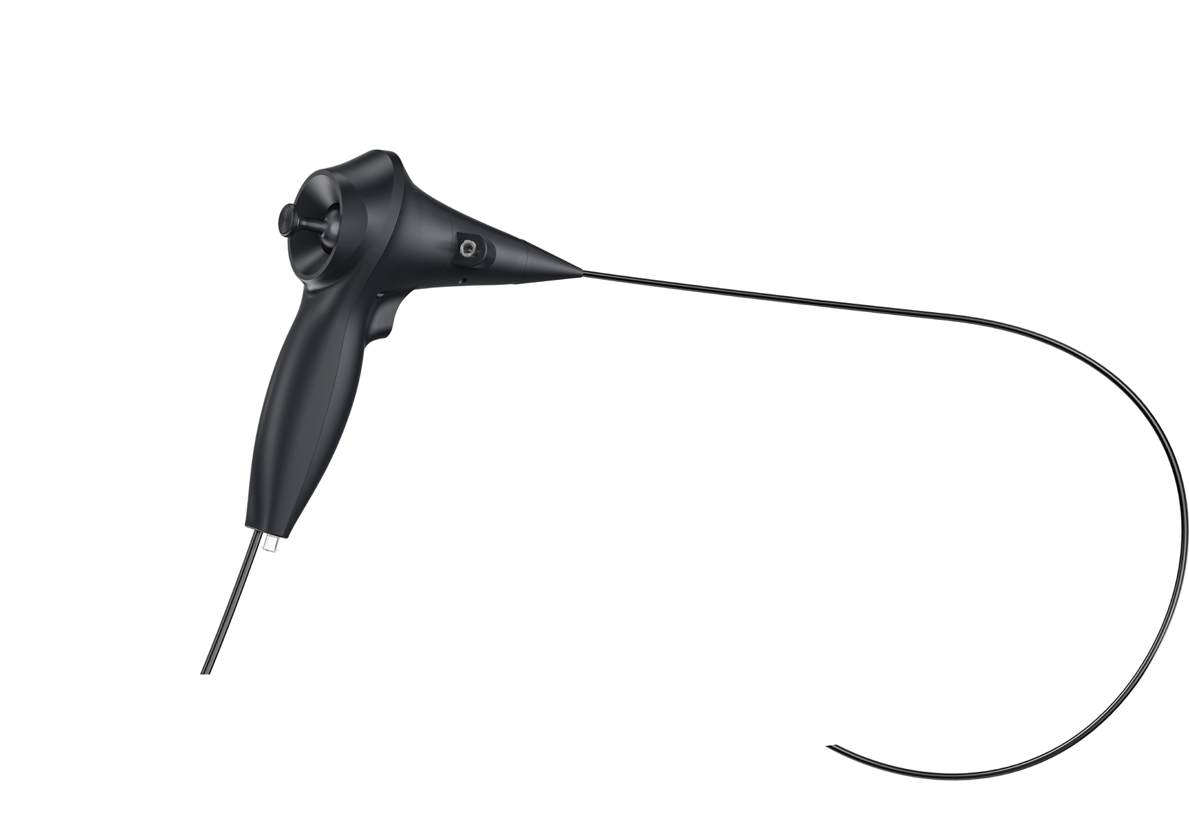
The insertion tube should provide appropriate reach without creating unnecessary handling burden. Tip deflection supports systematic inspection and controlled positioning within the stomach and accessible duodenum.
A usable working channel
The channel should be evaluated alongside the outer diameter. It determines compatibility with biopsy forceps, retrieval devices and other instruments, while also influencing suction capability.
Irrigation and suction
Fluid, mucus and gastric contents can obscure the field. An integrated irrigation and suction pathway helps the team restore visibility and manage fluid more efficiently during the examination.
Imaging and documentation
A stable image, practical display and image or video capture help the clinician document findings, support case review and maintain a useful clinical record.
LUMIVET CONFIGURATION EXAMPLE
A focused upper GI configuration.
| Insertion tube outer diameter | 7.0 mm |
| Working length | 1200 mm |
| Tip angulation | 130° all-direction bending |
| Working channel | 2.8 mm |
| Irrigation channel | Independent 1.0 mm channel |
| Supporting system | Irrigation and suction pump; compatible imaging workstation |
This example is based on the current Lumivet Veterinary Gastroscope configuration. Suitability must be assessed against patient size, anatomy and intended procedure. Product configurations may be updated.
A practical examination workflow.
- Define the clinical question. Review history, examination findings, laboratory results and diagnostic imaging to determine the goal and limitations of endoscopy.
- Prepare and check the system. Connect the gastroscope, imaging system and irrigation/suction equipment. Confirm image, angulation, airflow or fluid functions and compatible instruments before the patient enters the procedure.
- Prepare and monitor the patient. Follow the veterinary team’s protocol for fasting, anesthesia, positioning and physiologic monitoring.
- Insert and inspect systematically. Advance according to patient anatomy and the planned examination, maintaining deliberate orientation and documenting relevant landmarks.
- Manage the field. Use irrigation and suction when mucus, fluid or contents interfere with visualization.
- Sample or intervene when appropriate. Pass compatible instruments through the working channel for targeted mucosal sampling or selected procedures based on the clinical plan.
- Record the examination. Capture relevant images or video, document sampled sites and integrate the findings with the rest of the diagnostic work-up.
Clinical value—and important limits.
Upper GI endoscopy can provide direct visual information that radiography and ultrasonography cannot provide on their own. It can document visible mucosal changes and support targeted biopsy collection for histopathology.
It also has limits. Endoscopic biopsies are generally superficial mucosal samples, and the accessible gastrointestinal region is limited. Findings should therefore be interpreted alongside clinical signs, imaging, laboratory data and histopathology.
Foreign-body retrieval requires particularly careful selection. Proximal, accessible objects may be candidates for endoscopic retrieval, while distal, linear, perforating or otherwise complicated foreign bodies can require surgical management.
Build the setup around the case.
A useful upper GI endoscopy system brings scope handling, visualization, compatible instruments, irrigation, suction and documentation into one repeatable workflow. The right configuration depends on the patients you see and the procedures your team intends to perform.
Clinical references
- WSAVA Gastrointestinal Guidelines and standardized upper GI endoscopy resources
- Merck Veterinary Manual: Gastrointestinal Obstruction in Small Animals
- Merck Veterinary Manual: Chronic Enteropathies in Small Animals
This article is intended for veterinary professionals and general educational use. It does not replace patient-specific clinical judgment, training, product instructions or applicable professional standards.